Showing posts with label mouse. Show all posts

Friday, 5 July 2013

How to grow a liver in a petri dish and save lives

Liver donors are extremely rare - In 2011, 5,805 liver transplants were completed in the US and that same year, 2,938 people died waiting for one. Needless to say, an area that needs attention. In a first of a kind study by Takebe et al. published in Nature this week we may be a step closer to solving this problem.

The group has used human stem cells (I'll go into how towards the end) to create what they have dubbed 'liver buds', that is basically miniature human livers (about 4mm in diameter). They have then taken these liver buds and surgically implanted them into mice. The surgically implanted buds were able to substantially prolong the life of mice with liver failure! This is itself is a pretty amazing feat of science! Taking some human cells, growing a mini liver, throwing into an animal with liver failure and they live longer!

The liver buds not only prolonged the lives of these mice but incorporated themselves with the mouses blood system and continued to grow after the implantation. The buds took on many features of a fully-functional liver such as producing and sending out liver-specific proteins and signals. The figure below shows the implanted bud (in the dotted area) gaining blood vessels over time (shown by the increase in red in the dotted area).

Figure showing vascularization of liver buds (reproduced from Takebe et al. (2013))
These mini livers were the result of combining 3 types of human stem cell. First, pluripotent stem cells were converted into cells that are programmed to express liver specific genes (this is done by given the cells signals that trick them into thinking they're growing in a liver). Next, they combined these liver-specific cells with some endothelial cells (the kind of cells that make your blood vessels) and some mesenchymal stem cells (these can make bone, cartilage and fat). Over time, these groups of cells interacted and self-organized into the liver buds seen above. Below is an illustration of the process of creating the liver buds (panel A) but also a visualization of them forming over time (panel B). You can see how they start as a large smear and as time progresses they come together in an organized fashion.

Figure showing A) An illustration of the methodology of the experiment and B) showing the formation of the liver buds over time. (Reproduced from Takebe et al. (2013))
The figure below is what is called a "survival curve" which illustrates the rate of death of the mice in different conditions. Every vertical drop in the line is a mouse (or mice) dying. So a flat line means the group is surviving, a rapidly decreasing line shows they a dying.

There are only two lines you need to worry about. The dotted line at the very bottom is the group of mice that had the procedure done but didn't have a bud implanted (the control group), and the solid black line at the top is the group of mice that had the procedure done and did have the liver bud implanted.

Survival curve (reproduced from Takebe et al. (2013))
This shows that when the mice don't have the treatment, over a 30-day period, they die fairly rapidly. However, when they receive the bud implants, over the 30-day period, only ONE mouse died!

I don't know about you but I find that pretty amazing and VERY exciting. Of course as with most first of a kind studies, it needs to be replicated and further studies done but the implications this has for not only those with liver damage, but with any organ damage is huge and every exciting.

PS. For those of you who do not belong to an institution and cannot view the original article there is a summary here.

Friday, 21 June 2013

A viral infection of the eye that can cure blindness?

A very controversial area in science is gene therapy with the use of viruses (it really shouldn't be that controversial thou). I realise it sounds quite scary to be injected with an actual virus, but gene therapy using a virus as a tool is a remarkably elegant (and relatively safe) technique which deserves some time to clarify. A search for 'gene therapy' in Pubmed gives back just shy of 40,000 peer-reviewed scientific papers, so it is clearly an exciting, well-researched area of science.

Now, a bit of background. Viruses are sneaky little buggers and have evolved over time to sneak past your normal immune defences and infiltrate your cells, and from there trick the cell into making more of the virus. Normally this is a bad thing such is the case with HIV or the more common virus, the flu. So what happens, if we take away all the parts that allow a virus to make us sick and give it some specific DNA that a patient might be missing. Then the virus will go, sneak into the persons cells and trick the cell into making proteins from that DNA, but the proteins will be good for the patient and replace an otherwise missing protein.

There are many diseases that are caused by a mutation in the DNA which then means the protein isn't made in the patient, so if we could somehow deliver the protein (or at least the blueprints for the protein, DNA) into the cells then we could replace what was missing.

Gene therapy using an AAV virus. (Source: Wikipedia)

Gene therapy has been successfully and safely used to treat many diseases such as chylomicronemia, chronic lymphocytic leukemia, and multiple myeloma and a clever technique has recently been published to advance the use of this technique to treat certain types of blindness.

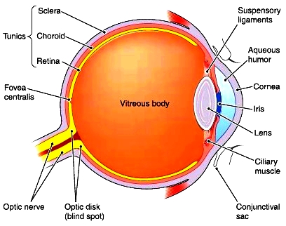
Inherited forms of retinal diseases afflict 1 in 3000 people worldwide and are primarily the result of mutations encoding proteins in the retina of the eye. Current gene therapy treatment for these diseases involves a needle piercing the eye and penetrating the retina itself to successfully deliver the virus. The diagram below shows just how far back that needle would have to go. The process requires hospitalization, general anaesthetic and runs a real risk of damaging the retina itself. This is because the special viruses usually used in gene therapy (called AAV) cannot get that far back into the eye normally and so need to be injected directly there.


Source: http://encyclopedia.lubopitko-bg.com

A group from Berkeley University has made use of the high rate of evolution in viruses (because they reproduce so quickly) and used directed evolution to "evolve" a virus that has the capabilities to get that far back in the eye. To do this, they took the standard AAV virus and injected it into the gel layer at the outer eye (the vitreous body in the diagram above), they then waited a while (a week) and collected the cells in the retina of the eye to see if any viruses made it that far back. There were some! So they let these few replicate into huge numbers again and repeated the process and repeated it and repeated it, each time only continuing with the viruses that somehow had the ability to reach the back of the eye, therefore selecting the viruses with the desired ability. At the end of all this they found 48 AAV viruses made into the back of the retina and 2 thirds of them were the same virus, they dubbed it 7m8.

To test if this virus could then be used to treat a disease, they used two mouse models of retinal degeneration caused by malfunctioning proteins in the retina and split each disease model into two groups. In one group they injected the virus, packaged with the DNA required to make the missing/malfunctioning protein, and the other group received the virus by itself without any extra DNA packaged into it. The mice with the 7m8 loaded with DNA had great vision improvements and a protein analysis proved that the retina was in fact producing the previously missing protein.

Immunostaining for RS1 protein (Dalkara et al. 2013)
The above figure is looking for the protein that is normally missing in the one of the diseases (RS1). Panel A shows a picture of the diseased retina and there is no RS1 protein there (you can tell from no greenness), panel C shows what a normal non-diseased retina should look like (lots of green) and in the middle (panel B) we have a diseased retina that had the injection of the virus with the DNA and as you can see it looks just the normal retina in panel C! Magic! Lots of green!

Now you might be thinking? So what? They still need an injection in the eye to get the virus in the first place, and you'd be right! However, injecting into the vitreous gel layer is a simple procedure, done at your local doctors practice, under local anaesthetic and runs very little risk of damaging the retina and this makes it a much more practical solution to those rendered blind from these diseases. Furthermore, this study, as a general concept, paves the way for developing more AAV viruses that are specialised to reach certain tissue or organs.

The study was published on the 12th June in Science Translational Medicine and can be viewed here.

Tuesday, 28 May 2013

First post ...

Hi there, this is the first post of my science blog. I personally think science is absolutely amazing. Humanities proudest achievement. Science stems from the word "scientia" which is latin for knowledge. It is the rational, logical process by which we, as intelligent apes, attempt to further understand the mechanics of this universe. In this light, I believe science is for sharing, so that all other intelligent apes can marvel at the beauty and complexity of this funny old world we live. However, to fully comprehend the published literature and to critically assess the findings and methodology is basically like learning another language for most people. So I thought that as a budding PhD student, constantly reading and critically assessing literature, I would write a weekly (to start with) blog post where I find a rad piece of science and serve it in an easily digestible piece of writing in order to share it with as many folks as possible with the goal of engaging the reader, not alienating the reader.

So to start with I have written a small piece on a paper that was published in the May edition of nature. It's a stunning piece of science at the crossroads between engineering, science and sheer awesomeness.

The authors (Chung et al.) have devised a methodology that allows an intact brain to become transparent. This isn't useful in itself, but when combined with other biochemical techniques that can stain particular types of cells, or a particular neuron or a specific region of the brain, this is where the magic happens. The authors created a 3-D image of a mouse brain seen here:


What gives the brain is "creamy" colour is the fat content of the cells, or the 'lipids'. Without going into the technical details, the technique (dubbed 'CLARITY') works by immersing the brain into a liquid over the course of a couple of days. During this time, the lipids are removed and replaced with a transparent 'hydrogel' that will maintain the structure of the brain but will remain clear. The authors were even able to apply the technique to a human brain that was preserved 6 years ago.
Figure taken from Chung et al. (2013)

The technique will allow scientists to trace connections throughout the brain and according to the authors even allow visualisation of two paired neurons either side of the synapse (the area where the two neurons connect and communicate).

As this technique becomes widespread and gets combined with other new and existing visualisation techniques, it will revolutionise how we study the brain, the most complex thing known to man.

Science. Is. Rad.